
A clear, proven way to diagnose vertebrogenic pain
To confirm that you have vertebrogenic pain, doctors use MRI to look for specific changes that occur with endplate inflammation, which are called Modic changes.
They're called “Modic changes” because in 1988, Dr. Michael Modic was the first to identify these visible changes to the endplates on an MRI. Think of Modic changes as scars that show up on imaging.
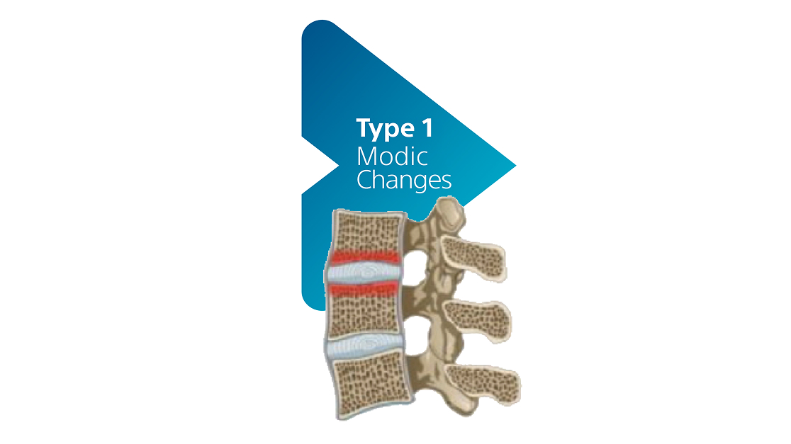
Type 1 Modic changes
Normal bone has a strong inner framework of connected structures. In the spaces between the structures, there is red bone marrow, which helps make blood cells.
With Type 1 Modic changes, a few things happen:
- Blood vessels start to develop in the bone/vertebrae
- Fluid buildup and inflammation occurs
- The bone’s inner structure stays the same—there’s no damage to the bone framework or marrow
In summary, Type 1 Modic changes means there's visible fluid buildup and inflammation in the vertebrae near the disc, but the bone itself isn’t broken.
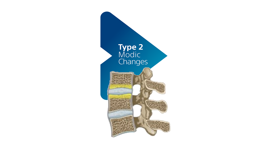
Type 2 Modic changes
Similar to Type 1 Modic changes, Type 2 Modic changes also have changes in bone marrow. However, in Type 2 Modic changes:
• The red marrow is replaced by fat
• This is the same fat found on bellies and hips
In summary, Type 2 Modic changes mean the vertebra bone near the disc has a fat buildup instead of healthy marrow. The bone structure is still there but is not as healthy.
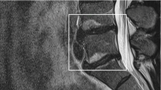
Photograph by Boston Scientific
MRI image showing Modic changes
For many years, there wasn’t a treatment for people whose MRIs showed Modic changes. For this reason, Modic changes can sometimes be overlooked on an MRI. Modic changes are now recognized as a clear and objective sign of vertebrogenic pain. It’s important to ask your doctor to look closely at your MRI for Modic changes.

Take the next step
Find a doctor
See if you may benefit from the procedure, and connect with an Intracept-trained physician near you.
Important Safety Information
Indications for use: The Intracept™ Intraosseous Nerve Ablation System is indicated for patients who have had chronic low back pain for at least six months, who have tried conservative care for at least six months, and whose MRI shows features consistent with Modic changes – indicating damage at the vertebral endplates has led to inflammation. Not every patient who meets these criteria is a candidate for the Intracept Procedure, however – in fact, there are specific characteristics indicating a patient should not be considered for the procedure.
Contraindications: Patients who are pregnant. Patients with weakened cardiac or pulmonary function, having an active implanted electronic medical device in the body (such as a pacemaker or defibrillator), being diagnosed with a systemic or local infection, or having an anatomy that could be damaged unintentionally while ablating the basivertebral nerve (based on your physicians’ clinical review). Patients who are skeletally immature – which generally means individuals under the age of 18 are not candidates.
Warnings: There are also certain risks and precautions regarding the procedure which you should be aware of before proceeding. With all medical procedures, there are risks and precautions associated with the procedure and the use of the device, talk with your doctor about what indicates, and contraindicates, certain patients for the Intracept Intraosseous Nerve Ablation System. For complete indications for use, contraindications, warnings, precautions, and side effects visit www.relievant.com/intracept/.
Caution: U.S. Federal law restricts this device to sale by or on the order of a physician.